Inverted T Wave On Ecg Rhythms

Ecg Normal And Inverted T Wave Waves Normal Ekg

Figure 2 Various T Wave Abnormalities Including T Wave Changes Related To Myocardial Ischemia Ecg Interpretation Qrs Complex P Wave

Inverted T Waves Are Seen In The Following Conditions Normal Finding In Children Persiste Emergency Medicine Hypertrophic Cardiomyopathy Intracranial Pressure

T Waves In Ischemia Hyperacute Inverted Negative Wellen S Sign De Winter S Sign Ecg Learning Ecg Interpretation Segmentation Normal Ecg

Pin By Jason Winter Ecg Educator On Ecg Ekg Study Memo Cards Cardiovascular System Ecg Rhythms Subarachnoid Hemorrhage

Figure 4 Secondary St T Changes Due To Lbbb Left Bundle Branch Block Lvh Left Ventricular Hypertrophy Rbbb R Ecg Interpretation Segmentation Normal Ecg

There are two patterns of t wave abnormality in wellens syndrome.
Inverted t wave on ecg rhythms.

Twitter Nursingstudents Nursing Tips Nurse Cardiac Nursing

Ecg In Myocardial Ischemia Ischemic Changes In The St Segment T Wave Ecg Learning Ecg Interpretation Normal Ecg Segmentation


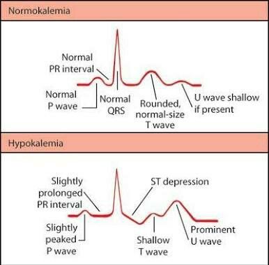
Visual For Ecg Changes For Different Electrolyte Imbalances Nursing School Survival Nursing School Notes Medical School Studying

Components Of The Ecg Strip Emergency Nursing Ecg Rhythms Cardiac Rhythms

T Waves In Ischemia Hyperacute Inverted Negative Wellen S Sign De Winter S Sign With Images Medical School Motivation Acute Coronary Syndrome Percutaneous Coronary Intervention

Medical Examinations Pulmonary Embolism Pulmonary Ekg

Junctional Rhythm Ecg Interpretation Ekg Cardiac Nursing

Ekg Injuries Used To Be Monitor Tech In An Icu Nurse Cardiac Nursing Nursing School

File T Wave Morphology Png Bestand

Ecg Presentation Slide 1 St Segment T Wave Segmentation Interactive Presentation Nursing Study
Pin On Ecg

Normal Ecg From 29 Y O Asymptomatic Athlete Sinus Bradycardia Early Repolarisation With St Elevation Arrows And Pe St Elevation Normal Ecg Cardiac Disease

Pin On Beautiful Body Art

Pin On Cardiac Rhythms

Figure 1 Discordance And Concordance Between Qrs Complex And St T Segment Ecg Interpretation Qrs Complex Normal Ecg

Ecg T Wave Checklist Ecg Interpretation Qrs Complex Interpretation

Junctional Rhythm If The Heart Rate Is Slow 40 55 Min The Qrs Complex Is Normal The P Waves Are Possibly Not Seen Then T Ekg Interpretation Arrythmias Ekg

Ekg Evolution Hyperacute T Waves Immediately 6 24hrs St Segment Elevation Immediately 1 6 Weeks Myocardial Infarction Cardiology Cardiac Nursing

Typical Ecg Abnormalities In Brugada Syndrom Brugada Syndrome St Elevation Abnormal

Click Through To Watch The Full Easy Ekg Interpretation Video Learn Ekg Interpretation In 10 Incredibly Easy Ste Nurse Nursing School Survival Cardiac Nursing
Pin On Medicine

Ekg Changes In Electrolyte Imbalances Nclex Quiz Electrolytes Imbalance Best Nursing Schools Pediatric Nursing

Heart Beat Ecg Tattoo Tattoos And Piercings Tattoos
Source : pinterest.com
