Inverted T Wave On Ekg

Ecg Normal And Inverted T Wave Waves Normal Ekg

Figure 2 Various T Wave Abnormalities Including T Wave Changes Related To Myocardial Ischemia Ecg Interpretation Qrs Complex P Wave

Inverted T Waves Are Seen In The Following Conditions Normal Finding In Children Persiste Emergency Medicine Hypertrophic Cardiomyopathy Intracranial Pressure

Ekg Evolution Hyperacute T Waves Immediately 6 24hrs St Segment Elevation Immediately 1 6 Weeks Myocardial Infarction Cardiology Cardiac Nursing

T Waves In Ischemia Hyperacute Inverted Negative Wellen S Sign De Winter S Sign With Images Medical School Motivation Acute Coronary Syndrome Percutaneous Coronary Intervention

T Waves In Ischemia Hyperacute Inverted Negative Wellen S Sign De Winter S Sign Wells Winter
The t wave should be concordant with the qrs complex meaning that a net positive qrs complex should be followed by a positive t wave and vice versa figure 17.
Inverted t wave on ekg.

Pin By Jason Winter Ecg Educator On Ecg Ekg Study Memo Cards Cardiovascular System Ecg Rhythms Subarachnoid Hemorrhage

Ekg Injuries Used To Be Monitor Tech In An Icu Nurse Cardiac Nursing Nursing School

Figure 4 Secondary St T Changes Due To Lbbb Left Bundle Branch Block Lvh Left Ventricular Hypertrophy Rbbb R Ecg Interpretation Segmentation Normal Ecg

Ecg In Myocardial Ischemia Ischemic Changes In The St Segment T Wave Ecg Learning Ecg Interpretation Normal Ecg Segmentation


Normal Ecg From 29 Y O Asymptomatic Athlete Sinus Bradycardia Early Repolarisation With St Elevation Arrows And Pe St Elevation Normal Ecg Cardiac Disease

Pin On Cardiac Rhythms

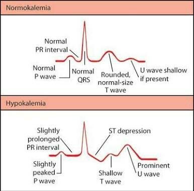
Visual For Ecg Changes For Different Electrolyte Imbalances Nursing School Survival Nursing School Notes Medical School Studying

Typical Ecg Abnormalities In Brugada Syndrom Brugada Syndrome St Elevation Abnormal

Pin On Nursing School Study Tips Nclex

Pin On Beautiful Body Art
Pin On Ecg

Dynamic T Wave Inversion Apparent Wellens Waves All Troponins Negative Unstable Angina Negativity Inversions Context

Rosh Review Syndrome Medical Mnemonics Cardiology

Pin On Medicine

2 646 Likes 5 Comments Medicohub Worldwide Medicohub Worldwide On Instagram Ecg Changes In 2020 Nursing School Survival Electrolytes Nursing Cardiac Nursing

Ecg Ekg Ischemia Injury Infarction Myocardial Ischemia Injury And Infarction Are The Different Types Of Damage Due To Ekg Cardiac Nursing Ekg Interpretation

Ecg Interpretation Characteristics Of The Normal Ecg P Wave Qrs Complex St Segment T Wave Ecg Learning Ecg Interpretation Qrs Complex Normal Ecg

The Ecg In Assessment Of Myocardial Reperfusion Ecg Learning Myocardial Infarction St Elevation Segmentation

Ecg Ekg And The Importance Of Potassium K In Short Very Important For Sustaining Cardiac Rhythm Nursing Tips Nurse Cardiac Nursing

B13176d9743fb734d93f7d125939912b S Wave Normal Ecg Jpg 494 395 Pulmonary Embolism Pulmonary Ekg

Ecg T Wave Checklist Ecg Interpretation Qrs Complex Interpretation

Pin By Ti Lo On Ekg Cardiology With Images Medical Mnemonics Ecg Interpretation St Elevation

Left Atrial Enlargement P Mitrale Right Atrial Enlargement P Pulmonale On Ecg With Images P Wave Ecg Interpretation Qrs Complex
Source : pinterest.com
